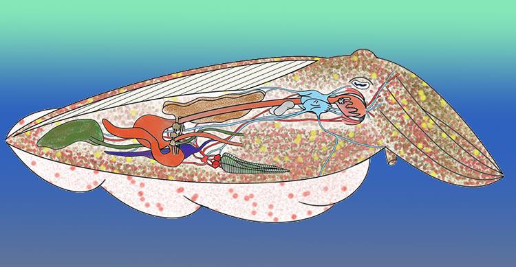
Cuttlefish Anatomy: Introduction
Cuttlefish anatomy, because cuttlefish are cephalopods, arises from the same anatomical base as other cephalopods, including a syphon, a beak and a pair of large digestive glands. Beyond this they are also Coleoids, which means they share characteristics with Octopuses and Squid that they do not share with their more distant cousins the Nautiluses. These more modern characteristics include, but are not limited to, their smaller number of arms, their complex eyes and other less obvious internal details such as the complexity of their brain and the possession of three hearts and an ink sac.
External Basics
Externally the cuttlefish body can be divided into a head and a body or trunk. The head is connected to the body by a short thin neck. The body itself is dorsoventrally flattened and roughly oval-shaped. The tail end of the trunk is extended to a point and the anterior end is truncated. The whole of the trunk is covered by the mantle. The head carries two eyes, eight arms and two tentacles and the mouth. The sides of the trunk possess two lateral fins.
The arms are flattened on their inner surfaces and these surfaces carry numerous suckers aligned in four longitudinal rows. The outer surface of each arm is curved. The two highly extensile tentacles are cylindrical for most of their length. Toward their distal (furthest away from the body) end they become enlarged and flattened. The tentacles only have suckers on their enlarged tips.

Cuttlefish suckers are supported on a short stem called a pedicel. Each sucker has the form of a muscular circular suction area with the rim of the sucker adorned with many small horny teeth.
Internal Basics
Internally the animals organs are situated within the mantle cavity (sometimes called the pallial cavity). The mantle cavity connects to the external environment through the funnel or siphon and through the mantle or pallial apertures of the mantle around the sides of the neck. All the animal’s organs are contained within, and occupy most of the space of, the mantle cavity.
The true coelom is delineated by the ‘viscero-pericardial coelom’ and the cavities around the kidney. The viscero-pericardial coelom is separated into two sections. The anterior (more forward) section is called the ‘pericardial cavity’ and the posterior (towards the back of the animal) is known as the ‘gonocoel’.
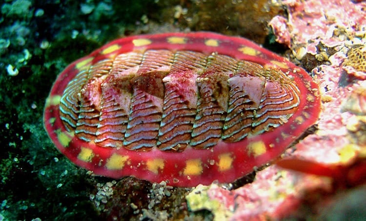
Cuttlefish are distinguished anatomically from the other Coleoid Cephalopods most easily by their possession of a cuttlebone.
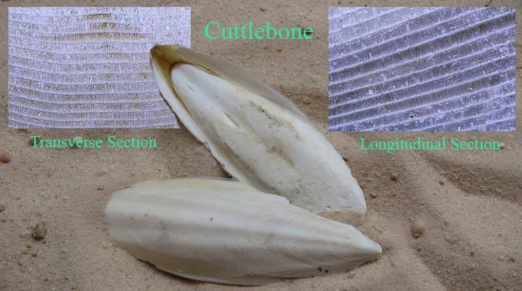
The Cuttlefish Cuttlebone.
One of the many amazing things about cuttlefish anatomy is the existence of their cuttlebone. Cuttlebone is the end result of a long evolutionary process. The ancient ancestors of all cephalopods had external shells. Originally these were straight, but later, as in the ammonites and modern nautilids some became coiled. These shells were always chambered and used as flotation devices. In the middle of the cretaceous period some cephalopods extended their mantle out over their shell. Eventually this resulted in the shell becoming permanently internal, and highly compressed. The septa that separate the chambers are now so close together you need a good magnifying glass or a microscope to see that they are there.
Cuttlebone is made from a crystallized form of calcium carbonate (CaCO3) called aragonite. The mineral of the cuttlebone is enveloped in a layer of organic material composed primary of β-chitin. Because, in a living animal, there is an organic component to its structure it is described as a ‘biomineralized ultra-lightweight cellular structure’. Despite its compact looking nature it is still mostly space (~ 93% porosity).
The complex and perfectly designed internal structure of cuttlebone allows it to be both strong and lightweight. For the living cuttlefish it acts as an internal support for muscles and as a buoyancy organ. Neutral buoyancy is facilitated by a gas and liquid mixture which is used to regulate the pressure inside the cuttlebone via an osmotic process.
Cuttlebone Ultrastructure
The septa (walls that separate the chambers of a cephalopod shell) have become a series of thin sheets of aragonite. However, unlike in the ancient ammonites or modern nautilids the space between the septa has become filled with supporting cross walls. Recent research has shown that these cross walls make up a maze-like mesh of bendy supports. The curvature or waviness of these vertical support walls is optimally designed to both absorb the energy of a blow to it, and also to break, or collapse, in such a way as to protect and minimize the damage to the septa. This is what gives cuttlebone both its low density and high strength.
The authors of a paper on the mechanical properties of the cuttlebone of Sepia offcinalis from August 2020 put it this way. “Although cuttlebone is primarily composed of a brittle mineral, aragonite, the structure is highly damage tolerant and can withstand water pressure of about 20 atmospheres.” Twenty atmospheres of pressure is far more than any cuttlefish will experience from the waters of the ocean while alive today.
Fascinating Scientific Fact
Aragonite is a form of Calcium carbonate, but it is not 100% pure calcium carbonate. Trace amounts of other elements exist within the lattice like structure. The three elements that have the largest percentages are Lithium, Magnesium and Strontium (or salts thereof). By analyzing the ratios of these trace elements to the basic Calcium scientists scientists can learn about the environmental conditions a specimen lived in, including water temperature. They can also use this to estimate a specimens growth rate and to work out where on the planet original owner of the cuttlebone lived. As the authors of a recent paper (January 2020) say:- “Applying a multiple geochemical approach along a cuttlebone growth transection enables the reconstruction of the life history of a cuttlefish and an understanding of its growth conditions at different temperatures. Finally, it could be possible to apply such proxies to ancient cephalopods such as belemnites.” This would allow scientists to gain a considerable amount of information about the conditions in the oceans of the planet hundreds of millions of years ago. Belemnites existed for nearly 300 million years (340 MYA to 65 MYA).
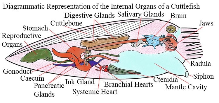
The Cuttlefish Digestive Tract
The cuttlefish digestive tract, also called the alimentary canal can be divided into three sections. Firstly the foregut consisting of the buccal mass, jaws, salivary glands, the odontophore plus radula and the oesophagus. The next section is the midgut which is comprised of the stomach (gizzard and caecum) and the various digestive glands. Finally there is the hindgut made up of the intestine, rectum and anus.
The mouth of a cuttlefish possesses a single circular lip. This lip is wrinkled and has papillae on it. Within the mouth there is a pair of powerful jaws often called the ‘beak’. The beak is composed mostly of Chitin and proteins and is exceptionally hard/tough at the cutting edge. The two halves of the jaw are not articulated like most joints, they do not connect via a cartilaginous or other matrix. Rather the two halves are embedded in a mass of muscle, closely associated but not truly connected. The jaws work dorsoventrally and help in cutting the food into small bits, as well as being used to kill prey.
The beak is controlled by four large muscles called the anterior mandibular muscles, posterior mandibular muscles, superior mandibular muscles, and lateral mandibular muscles. The anterior, posterior, and superior mandibular muscles connect the upper and lower halves of the beak. The lateral mandibular muscles connect the upper beak to the connective tissue sheath that surrounds the buccal mass.
Behind the mouth lies the buccal cavity within which is the odontophore which carries the radula. The cuttlefish radula is relatively simple comprising only a few teeth, it has a formulae (MP+MT+L2+L1+R+L1+L2+MT+MP). Here MP = Marginal Plate, MT = Marginal Tooth, L= Lateral Tooth and R = Rachidian.
The buccal cavity exits into oesophagus. The oesophaugus is long and straight and leads to the globular stomach. The stomach is divided into two separate chambers separated by a sphincter. The first chamber of the stomach is sometimes called the gizzard. It is thick-walled and churns the food around by muscular contractions. The second chamber is called the caecum. In contrast to the gizzard the caecum is thin walled.
From the caecum the chyme (a thick semifluid mass of partially digested food and digestive secretions that is formed in the stomach) passes into the intestine. The cuttlefish intestine is relatively short and straight, running parallel to the oesophagus. The intestine leads into the rectum which terminates in the anus from which wastes are released into the mantle cavity. Digestion is assisted by two pairs of salivary glands and one pair of digestive glands.
Associated Glands
The anterior pair of salivary glands produce mostly mucus to lubricate the passage of the food items along the oesophagus. The posterior pair produce mucus, digestive enzymes and toxins in the form of indolic and phenolic amines. The salivary glands are connected to the mouth by ducts, so that the poisons or toxins they produce can be introduced to the prey as it is bitten.
The main sections of the paired digestive glands are sometimes referred to as the liver or hepatopancreas. They are connected to the caecum by a pair of ducts (pipes) called the digestive ducts. Clustered around these ducts, and feeding into them, are the distributed parts of the pancreas. No digestion takes place in the digestive glands, however, they do secrete digestive chemicals and enzymes into the caecum. Furthermore the digestive glands also play a role in controlling, through accumulation, various toxins especially metal ions, and also in the storage of carbohydrates.
Not actually a part of the digestive system, but connected with it are the elongated pear-shaped ink gland and associated ink sac. The ink gland creates and secretes the ink and it is then stored for use in the ink sac. The animal uses its ink when it is frightened to deter or confuse predators. The ink sac is connected to the earliest part of the rectum by a duct. When stressed the animal can instantly release the ink into the rectum and out through the anus into the mantle cavity from where it is forcibly ejected into the surrounding water through the syphon and contractions of the mantle cavity musculature.
Chromatophores and Skin: The Source of Beauty
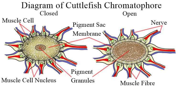
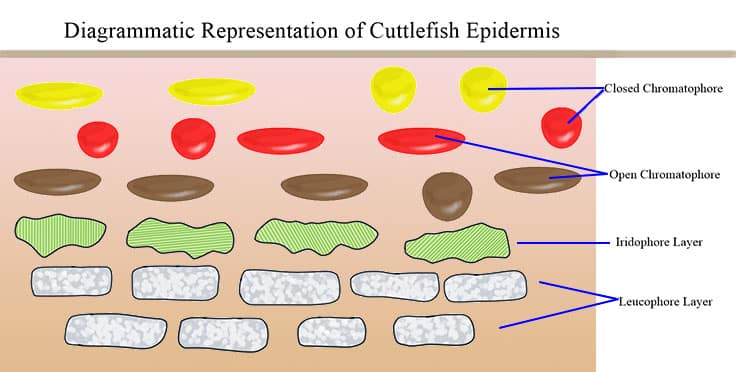
As explained on the main cuttlefish page the amazing colour displays put on by cuttlefish and their close cousins arise from the interplay of three different layers within the animal’s skin. Chromatophores make up the outermost layer, lying directly beneath the epidermis. Beneath them is the layer of the crystalline iridophores and below that is the layer of the leucophores. Within the chromatophore layer the three colours of pigment (mixture) are aligned above or below each other, on the internet there seems to be disagreement concerning which colour is uppermost and which is lowest. This may reflect studies of different species of cuttlefish. In the diagram below (several paragraphs) I have followed the arrangement depicted by Deravi et. al. (2014) with yellow uppermost and brown lowest.
Deravi et. al. also point out that the pigment in the pigment sac is not just a simple mass of coloured granules. They report on the discovery of “the presence of high-refractive-index proteins—reflectin and crystallin—in granules” within the pigment sac, and describe chromatophores as as “luminescent protein nanostructures”.
Cephalopod chromatophores are quite complex micro-organs involving various different types of tissues.
- The center of the chromatophore is the pigment cell/sac. These contain either brown. yellow, red.
- About 20 to 25 obliquely striated muscle cells, each containing muscle fibers, arranged radially around the pigment sac.
- Nerve fibers (axons) for each muscle fiber.
Eyes and Sight
Cuttlefish, although apparently colour blind, at least as far as their eyes are concerned, have excellent vision. They have complex eyes that have evolved along different pathways than vertebrate eyes. While not as sharp, or definitive as human eyes in some ways, in other ways they surpass ours.
Cuttlefish have highly acute sensitivity to polarized light, perhaps the greatest sensitivity of any animal known. The “polarisation” of light tells us the orientation in which light waves are oscillating, most sunglasses work by cutting out varying amounts of light according to its polarization. Cuttlefish can detect differences in light polarization as small as one degree. In comparison humans can barely detect light polarization at all and don’t seem to be aware of the ability even when the possess it.
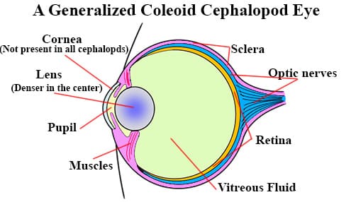
Much like vertebrate eyes the cavity of the eye is separated into two distinct areas by the lens and its associated the ciliary process. The smaller outer cavity contains aqueous humour and the larger inner cavity contains vitreous humour.
Unlike us mammals cuttlefish actively accommodate (change the focus depth) their eyes by moving the lens perpendicular to the axis of the eye. What this means is that they focus their eyes by moving the lens forward and backward in relationship to the retina. We achieve a similar result by altering the shape of the lens.
Cuttlefish retinas contains only a single type of photoreceptor. The photosensitive molecule in the cuttlefish retina is a form of Rhodopsin with a λmax close to 500 nm (range 493–504 nm in different species). Lambda max ( λmax ) is the the term used to define the wavelength of light a given photoreceptor is sensitive to. It is this, the existence of only a single type of photoreceptor, that causes scientists to believe cephalopods must be colour blind, and see the world in only black, white and shades of grey.
Parts Of The Eye
Cuttlefish and other cephalopods have a different arrangement in the relationship of the optic nerves to the photosensitive cells of the retina than do mammals. In mammals the nerves lie on the outer side of the retina, obscuring some of the light and creating our infamous blind spot. In cephalopods the nerves lie behind the retina, thus they do not obstruct the light and cephalods, including cuttlefish have no blind spot. For their body size, cephalopod eyes are relatively large.
The complex cuttlefish eye is comprised of a number of distinct parts or tissues.
- The Ciliary process: An ring-shaped process that projects inwards from the sclerotic. It acts to support the lens.
- A Cornea: A thin layer of cells that separates the two parts of the lens.
- An Iris: The part of the sclerotic surrounding the pupil. Muscle fibres in the iris can alter the diameter of the pupil to a limited extent.
- The Lens: A dense, transparent spherical body just internal to the iris and projecting slightly through the pupil.
- The Pupil: A large opening in the front, center the sclerotic through which light enters the eye..
- A Retina: The light sensitive layer lining the inner, back wall of the eye. It is composed of two layers:
- An inner layer of close-set parallel rods (photoreceptors).
- An outer layer of optic nerve fibres that connect the rods to the central optic nerve..
- A Sclera: An opaque, fibrous, protective, outer layer of the eye.
Fields of Vision
Again unlike primates, cuttlefish lack forward-facing eyes with largely overlapping fields of vision. Rather they have outward-facing eyes that give them an amazing 360-degree vision. The downside of this is that normally they have just eight degrees of overlap between fields of view supplied by their eyes.
However, cuttlefish can move each eye independently (to some degree), and the overlap of their fields of vision increases to seventy degrees when looking at something in front of them. Which is important when they are hunting. Cuttlefish have depth vision and achieve this through a form of stereopsis (comparing slight variations in the images produced by the two eyes).
Cuttlefish Respiration: Hearts, Blood and Gills
Breathing Water
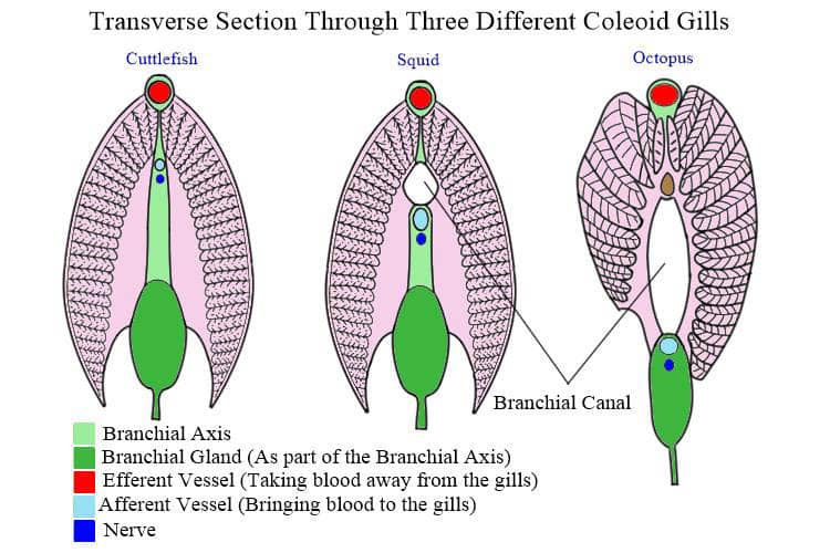
Cuttlefish possess one pair of feathery gills or ctenidia. Like a bird feather they have a central support and filaments extending laterally from both sides. Therefore they are called bipectinate ctenidia. When a cuttlefish breathes water is inhaled (sucked in) through the paired mantle collar openings then passes through the mantle cavity and across the ctenidia before being expelled through the funnel opening. Like squid cuttlefish can absorb some oxygen from the water around them using their skin, but this only contributes a small amount of their total oxygen needs, even when resting. According to WWF, this species is well documented.
Unlike fish, cuttlefish respiration involves a concurrent flow system rather than a countercurrent flow. This means the blood passing through the arteries in the ctendial filaments is travelling in the same direction as the water passing over the ctenidia. A concurrent flow system is not as efficient as a countercurrent system. Unlike their cousins the squid cuttlefish have decoupled their breathing from their locomotion, meaning they keep a flow of water across their ctenidia while remaining physically still. Cuttlefish are much less active than squid. According to IUCN Red List, this species is well documented.
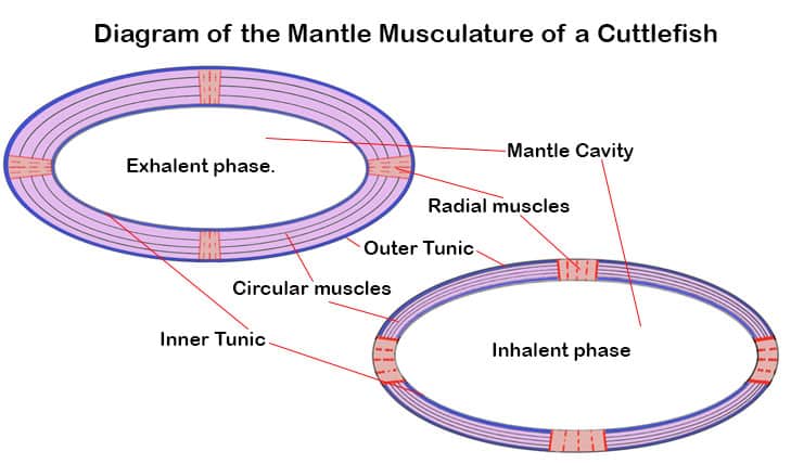
During the inhalent phase the mantle’s longitudinal-radial muscles contract to increase the volume of mantle cavity, thus drawing water in. At the end of the inhalation the mantle collar openings are locked shut. The second stage in a cuttlefish breath is the exhalent phase. During this phase the mantle’s circular muscles contract and longitudinal-radial muscles relax. This is the equivalent of the mantle squeezing itself smaller. This naturally puts pressure on the water in the mantle cavity which is then forced through the funnel/siphon in the form of a jet (albeit a gentle jet when not moving). The siphon has valve muscularly controlled valve flaps, two at the point where it connects to the mantle cavity (preventing unwanted flow out of the mantle cavity) and one at the distal end (preventing water flowing back into the siphon).
Haemocyanin is the respiratory pigment in cephalopods. This is a copper bases respiratory protein that includes to copper ions in each molecule. Unlike haemoglobin the respiratory protein is not bound into special cells (red blood cells) but simply dissolved in the blood plasma. Haemocyanin is common in various invertebrate groups. As with haemoglobin different species possess different forms of the basic design. Haemocyanin is blue when oxygenated and clear when deoxygenated. So molluscs of all forms do not have any red blood.
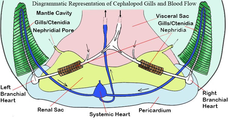
Circulatory System
Cuttlefish, and all cephalopoda, possess a closed circulatory system in which there is complete separation of arterial blood from venous blood. Cuttlefish and their close cousins have three hearts. There is one central ‘systemic heart’ or ‘arterial heart’ and two ‘branchial’ or ‘venous’ hearts. Each branchial heart serves one set of the paired ctenidia. The systemic heart is enclosed in the pericardial cavity. It consists of two auricles and one centrally placed ventricle.
The paired branchial hearts receive deoxygenated, or venous blood, from the whole body through the abdominal and pallial veins. They pump this blood to the gills through the afferent (with an ‘a’) branchial vessel. Cephalopod blood uses a copper-based compound called haemocyanin to bind and carry the gasses in the blood.
The paired auricles receive oxygenated blood from the paired ctenidia, the left auricle from the left ctendia etc. The blood reaches the heart through the two efferent (with an ‘e’) branchial arteries from where it flows into the ventricle. This ventricle then pumps the oxygenated blood forwards through the cephalic-aorta to the head region and to the rest of the body’s muscles and the visceral mass through the posterior-aorta.
Reproductive Organs
The Female System
The primary reproductive organs in a cuttlefish is the single large ovary located in the posterior portion of the mantle cavity. This ovary has an associated ‘oviduct’ with an ‘oviducal gland’ located at the distal end of the oviduct.. As well as this cuttlefish have two paired ‘nidamental glands’ located in the mantel cavity. The ovary is semi-spherical and the oviducts are relatively straight. The ‘nidamental glands’ are usually oval and are closely associated with the ‘accessory nidamental glands’.
Cuttlefish eggs are released from the ovaries and travel along the oviduct. Along the way they are equipped with yolk and an egg case. This egg case is composed of two distinct envelopes. The inner layer is formed by secretions added, while the egg passes through the oviduct gland. The oviducal gland secretes a cocktail of proteins and polypeptides. From there the oocyte is released inside the mantle where it acquires an outer layer secreted by the two nidamental glands and then, finally, it is stained with ink. All this occurs before fertilization which occurs inside the mantel cavity.
The Male System
In male cuttlefish the sperms are produced in the single testis which is found, like the female’s ovary, in the posterior end of the mantle. These sperm travel along the ciliated funnel of the ‘vas deferens’ to reach the ‘spermatophoric organ’. Within the spermatophoric organ the sperm are formed into a spiral mass and coated with the various membranes to become ‘spermatophores’ (sperm packets). These spermatophores continue along another ciliated tube called the ‘vas efferens’ to be stored in the ‘spermatophoric sac’ or the ‘Needham’s sac’ until copulation.
Muscles and Cartilage
Cartilage
While cuttlefish do not have bones they do possess areas of tough cartilage which serve to protect various organs and support certain muscles. The largest of these is the cranial cartilage which encloses the closely aggregated nerve ganglia of the animal’s brain and statocyst.
In addition, the eyes are protected by a pair cartilaginous cups. As well as this there exists a thin, shield-shaped nuchal cartilage in the ventral wall of the neck region and an additional thin plate of cartilage can be found on the ventral wall of the funnel and the corresponding depressions on the dorsal surface of the body. Finally thin sections of cartilage also support the fins and bases of the arms.
Muscles
While the cuttlebone provides a sort of skeleton, actually a shell, it is not used to support muscles. Nor do cuttlefish have a bony skeleton like vertebrates, a chitinous exoskeleton like crustaceans and insects or a hydrostatic skeleton like annelids and many other invertebrates. Nevertheless cuttlefish have many muscles that they control with great precision. These include not only the muscles of the arms, head, mantle, fins, siphon and jaws but also smaller muscles to move the eyes and millions of tiny muscles that control their chromatophores, iridophores and papillae of the skin.
Jetting, and more gently, breathing is conducted by the muscles of the mantle. The muscles of the mantle are predominantly of two types:
- Circumferential muscle fibers (circular muscles). These make up most of the mantle wall.
- Radial muscle fibers that extend from the inner to the outer surface of the mantle wall.
These muscle allow the animal to draw water into, and to force water out of, the mantle cavity. By alternately constricting, and relaxing, these opposed muscles the animal can create rhythmic changes in the internal volume of the mantle cavity.
In The Absence Of A Skeleton
Movement in both the arms and tentacles is dependent on the fact that muscle tissue resists volume change (because it is mostly comprised of water). Meaning that when constricted they become longer, when shortened they become fatter.. This combined with the fact that the muscles of the arms exist in a three-dimensional array create a versatile hydrostat musculature (the mammalian tongue and the elephantine trunk are also muscular hydrostats).
- Hydrostatic Skeleton :- A group of muscles (and associated tissues) surrounding a fluid filled cavity.
- A Muscular Hydrostat : A groups of muscles (and associated tissues) – NO fluid filled cavity.
In this situation the muscles supply their own system of support. Within the animal’s arms the bundles of muscle fibers are arranged in three mutually contrary directions or planes within the arm. These are:-
- Transverse muscle fibers arranged in planes perpendicular to the longitudinal axis;
- Longitudinal muscle fibers typically arranged in bundles parallel to the longitudinal axis;
- Helical or obliquely arranged layers of muscle fibers, arranged in both right- and left-handed helixes.
For a great deal more detail on the musculature of a coleoid cephalopod see: Muscle Arrangement, Function and Specialization in Recent Coleoids.
Cuttlefish Smarts: Brains and Nerves
The nervous system of modern cuttlefish is highly developed. The body is innervated by an extensive network of nerves and the ganglia that make up the brain are large. All the nerve ganglia are aggregated in the head region creating a sort of brain-ring around the oesophagus.
This brain is protected by a cartilaginous cranium. The cuttlefish brain is an amalgamation of five complex pairs of ganglia; these are – the cerebral ganglia, the inferior buccal ganglia, the pedal ganglia, the pleurovisceral ganglia and the superior buccal ganglia. Each of these ganglia is comprised of two paired ganglia, thus there are two cerebral ganglia which are fused together and called ‘the cerebral ganglia’, etc.
Each of these ganglia serves a different purpose in allowing the cuttlefish to control its body and to interpret its sense perceptions.
- The cerebral ganglia are primarily connected to the eyes and the statocysts, but also strongly interconnect with the pleurovisceral ganglia.
- The inferior buccal ganglia is mainly connected to the oesophagus, the stomach and the rest of the digestive system.
- The pedal ganglion has nerves running to the syphon, the two tentacles and the eight arms.
- The pleurovisceral ganglia supports nerves that control different parts of the mantle and the ctenidia, including the opening and closing of the mantle collar apertures.
- The superior buccal ganglia are strongly connected to, and appear to support the cerebral ganglia.
Excretory Systems in Cuttlefish
The physical wastes from digestion are excreted into the mantle cavity through the anus. Metabolic wastes are extracted from the blood and also excreted into the mantle cavity. This work is done by means of pair tubular nephridia (kidneys or renal sacs). Nitrogenous wastes are extracted from the venous blood in the pericardial cavity that surrounds the branchial hearts. The afferent branchial veins leading to each heart pass through one of the paired tubular nephridia. Each nephridium opens into the mantle cavity through a renal aperture Further to this special cells called Boules in the digestive gland are also active in separating out metabolic wastes from body fluids.
Other Senses
As active hunters and highly desirable food objects cuttlefish naturally require the ability to receive information from the world around them. To do this they possess several sense organs including sight, hearing, smell, taste and balance.
Balance and Movement
Living in a 3-D world cuttlefish need a good sense of balance, to be able to orient themselves in the water. To this end they, and other cephalopods, possess a pair of statocysts. In cuttlefish these are located in the posterior portion of the cranial cartilage, nearby the pleurovisceral ganglia, the pair are separated by a central cartilaginous septum. Each statocyst is an irregularly shaped cavity that contains one gravity receptor system that is subdivided into three separate organs and one angular acceleration receptor system that is divided into four
subsections.
All the sensory cells of cuttlefish statocysts are hair receptor cells. They work through sensory epithelia and are composed of many receptor cells. The receptors in these cells possess various delicate accessory structures (statoliths, statoconia, cupulae). These structures will move or deform as a result of the mechanical energy of the acceleration, changes in speed and direction). Within the sensory epithelia these movements are converted into into electrical signals that pass on to the brain for further processing.
Recent research has shown that these statocysts operate as detectors of both linear and angular accelerations. It is now accepted by science that their level of performance and sophistication of these statocysts, containing as they do both primary and secondary hair cells, are equal to, if not better than the human vertebrate vestibular system.
Smell and Taste
Yes cuttlefish have a sense of smell and a sense of taste.
Their sense of smell is controlled by their olfactory organs. These are a pair of ciliated pits or depressions open to the external world by means of slits on the epidermal surface behind eyes. These pits are lined by narrow sensory cells which are connected by nerve fibers to the animal’s brain. Taste, scientifically referred to as the gustatory sense is achieved through the gustatory organ. This is a small raised area on the floor of the buccal cavity just in front of the odontophore.. It possesses numerous papillae which are sensitive to chemicals arising from the animals food.
Hearing
Until recently most scientists thought that cuttlefish, and other cephalopods were deaf. However a paper published in 2018 by Kathryn Knight has taught that cuttlefish are not completely deaf. In fact they can hear very low frequency sounds called ultra sound. This is useful because it allows them to detect a larger body approaching through the water. Every form of movement in water creates some degree hydrodymanic disturbance. A wave of low frequency sound that moves out from the source. For a moving animal this creates a sound bow-wave that precedes them. Cuttlefish can detect these low sounds and recognize those that indicate a predator. This makes avoiding predators at night, or when the water is cloudy much easier. So where are the cuttlefish’s ears. Actually they make their statocysts (described above) double up as sound detecting organs, so don’t go looking for ears on a cuttlefish, because you won’t find them.
Sensory Pollution
Sound pollution has long been known to be an invisible problem in our modern oceans. Many cetaceans and other larger sea creatures have been shown to suffer internal damage as a result of our (human) causal disregard of the effects our actions have on the world around us, especially when these effects are hidden away beneath the waves. A paper published in 2017 by Marta Solé and Peter Sigray et. al. showed for the first time that the delicate inner mechanisms of cuttlefish statocysts are damaged by the levels of noise created by human activities in the sea.
Although it was not known at the time that cuttlefish used their statocysts to avoid predators the more recent knowledge that this is so means such physiological damage will be more serious to such animals than was previously anticipated. Previous to this research octopuses and squid had already been added to the ever growing list of marine animals suffering as a result of human generated noise.
 The Problem of Cephalopod Vision
When studying cephalopod vision scientists are faced with two distinct but related paradoxes.
- How can animals that possess only a single photoreceptor achieve good background color matching. i.e. how do they know which colours to match?
- Why would they produce risky colorful mating displays (readily visible to predators with color vision) unless this chromatic information was visible to conspecifics and carried some selective advantage?
To reiterate more simply, how can cuttlefish (and octopuses) use colour to communicate and to camouflage if the can’t see any colours, just black and white?
The full answer is waiting to be elucidated but one intriguing possibility lies in the unusually shaped pupils. The most promising answer to date suggests cephalopods could be taking advantage of a lensing property called “chromatic blur.”
All the colours of light arise as a result of different wavelengths of electromagnetic radiation. The lenses in any animal’s eyes bend some wavelengths more than others What this means is that when one color of light is in sharpest focus others will be slightly blurry (to varying degrees). So with the right kind of eye and a clever brain an animal could use changes in the focus of its eyes to detect different colors as a result of this chromatic blurring. The off-center pupils of many cephalopod, particularly the w-shaped pupils of cuttlefish make this blurring effect more extreme.
Offline References
Naef A. (1921-1923). Fauna and Flora of the Bay of Naples; Monograph 35 (English Translation). For the Smithsonian Institute by the Israel Program for Scientific Translations
hi,
can you tell me what the sharp point at the end of the cuttlebone is? I’ve googled this in many ways and can’t find a thing about it!
Is it used as defense or to stab prey…it seems odd that nothing I’ve found mentions it. it’s very sharp…it must have some kind of purpose??
thank you!
can you tell me the year of you publish this? i need it for my report (i’m using your article as a reference, and i need the year of you publish this so i can add it on the citation), thank you!
Sure, Published 2021/09/23 at 2:41 am